You can download a PDF version of this page here: Teddy’s Pigmentary Keratitis
I can see clearly now ……..
But I couldn’t until I had treatment for Pigmentary Keratitis
Read my story …
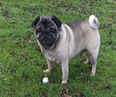
This is the story of a pug surrendered to PDWRA for rehoming.
As part of PDWRA procedures he was taken to the fosterer’s vet for a health check. He was generally healthy but had limited vision. This led to an eye examination but the vet was unable to see into his eyes due to a covering of pigmentation.
With this type of condition the vet recommended referral to a specialist ophthalmic veterinary practice. This was done and he was seen by a specialist.
Diagnosis
The ophthalmologist diagnosed advanced Pigmentary Keratitis (PK) in both eyes.
In simple terms a thin film of tissue (pigmentation) was growing over the outer surface of the eyes. As the pigmentation progresses light is prevented from reaching the parts of the eye that enable vision. If the growth of the pigment is not stopped (or removed) vision will continue to deteriorate. Additionally there was inflammation caused by deformed lower eye lids turning in resulting in the eye lashes rubbing on the eye.
(It’s like being in a room at night with no light. No light no sight.)
After discussion with the ophthalmologist it was decided the pigmentation had progressed too far to achieve any significant improvement using suppressive drops, and surgery would be the best option to improve his vision and quality of life.
Summary of Surgical Report
The majority of the pigmentation was successfully removed by laser. As a result the ophthalmologist was optimistic that the vision from both eyes would improve. In effect what the surgery does is like scraping the ice off a car windscreen (surface of eye) on a frosty morning but to keep the screen clear wipers and antifreeze are required. For PK patients post-op drops are used to keep the cornea clear.
In addition plastic surgery was performed to restructure the eye lids to reduce the aperture size and eliminate any rolling in of the lower eyelids to prevent irritation from the eye lashes.
Post-operative care for first 10 – 14 days
Notes:
- There are small sutures in the corner of the eyes which will dissolve over several weeks.
- Expect some swelling around the eyelids.
- There may be some bloody discharge around the incisions over the next 24 hours.
- Bathe away any discharge with cooled boiled water and pat dry with tissue.
- Keep collar on at all times.
Medication:
- 4 types of drops to both eyes representing 13 occasions daily.
- 2 types of tablets once daily with food.
Proper management of immediate post op care is essential and requires significant time and patience.
Post-op check 13 days after operation
This confirmed the laser surgery had significantly reduced the pigmentation and improved vision. This allowed medication to be reduced to a more manageable level:
- One drop both eyes twice daily.
- One drop both eyes four times daily for ten days, then stop.
- One drop both eyes four times daily (apply last).
The next check was scheduled for 3 months’ time, sooner if any concerns arose.
3 month post op examination
On re-examination both eyes were open, comfortable and had evidence of good vision. The left corneal pigmentation continues to disperse well, with a clear view of the posterior segment* permitted. The right corneal pigment has cleared well in the lateral 2/5 of the cornea but the central and medial cornea remains pigmented**. This should continue to clear with continued use of the Tacrolimus.
The next check was scheduled for 6 months’ time.
Explanatory notes:
* The posterior segment is the back two-thirds of the eye that includes all the optical structures that enable vision. In cases of advanced PK this part of the eye is not visible due to pigmentation and critically prevents light getting to the optic nerve causing loss of vision.
**The right eye had not cleared as well as the left due to an ulcer being present at the time of surgery.
What is PK?
PK is a condition that results in progressive inflammation and pigment deposition over time on the corneal tissue (the clear surface of the eye). The condition can develop at any age and has been seen in puppies as young as 12 weeks of age.
It is known that flat faced breeds e.g. Pugs, French bulldogs, Bulldogs, Pekinese, Shih Tzu, and Lhasa Apso are predisposed to this condition. Thus owners of these breeds can take preventative steps to stop the disease developing, and in cases where the condition has developed to the stage where vision is impaired treatments are available to restore good quality vision.
There is no treatment available at this time to completely reverse this disease but there is medication which is known to suppress the progression.
What causes the condition to develop?
The cause of PK is not yet fully understood, but there is a strong correlation between genetic makeup, trauma to the eye (such as chronic irritation, corneal ulcers, or scarring), and the development of pigmentation. Chronic irritation is usually caused by a congenital defect of the eyelids that results in the eyelids rolling inwards making the eye lashes rub against the eye ball.
Readers will find the following web site useful to get a further understanding of this disease, its causes and treatment:
www.dogquality.com/blogs/senior-dog-blog/13922319-corneal-pigmentation-in-brachycephalic-dogs
Summary of what is known about PK
The good news is that:
- A lot is now known about PK and how it can be treated.
- It is known that the pigmentation starts from “small beginnings” and progresses very slowly so it may go unnoticed for a long time which means the pigmentation is more difficult to deal with.
- It is known that a major contributor to the cause of the disease is physical damage to the outer surface of the eye, e.g. ulcers, eyelids and skin folds in the vicinity of the eyes irritating the surface through contact, and dry eye.
- It is known there are medications which suppress the progress of pigmentation.
- It is known surgery will significantly help severe established conditions by resetting the baseline condition and allow medication to be more successful.
The not so good news is that pugs are a vulnerable breed. BUT KNOWING THIS IS A BIG PART OF THE BATTLE AGAINST THIS CONDITION.
What can Owners do?
The essential message for owners is “CATCH IT EARLY”. PK is a progressive disease and when caught in the early stages is much more manageable. Don’t wait until one day you suddenly notice your pug is not seeing things very well. When that happens it’s likely that PK may be at an advanced stage.
- It is recommended that pugs should be checked for the presence of PK every 6-12 months.
- If you have a reason to visit your vet soon ask for an eye examination for the presence of pigmentation.
- If you don’t have an early visit planned make an appointment.
- If the whites of the eyes look red and sore make an appointment.
A general practice vet should be able to see the early signs and definitely see established pigmentation with their standard equipment. If any pigment is found and you would like a specialist’s view , your vet can arrange a referral to a specialist ophthalmic practice. Specialist ophthalmic practices only work on a referral basis.